Mesothelioma is a form of cancer that is almost always caused by previous exposure to asbestos. In this disease, malignant cells develop in the mesothelium, a protective lining that covers most of the body's internal organs. Its most common site is the pleura (outer lining of the lungs and chest cavity), but it may also occur in the peritoneum (the lining of the abdominal cavity) or the pericardium (a sac that surrounds the heart).
Most people who develop mesothelioma have worked on jobs where they inhaled asbestos particles, or they have been exposed to asbestos dust and fibre in other ways, such as by washing the clothes of a family member who worked with asbestos. Unlike lung cancer, there is no association between mesothelioma and smoking.[1] Compensation via asbestos funds or lawsuits is an important issue in mesothelioma (see asbestos and the law).
The symptoms of mesothelioma include shortness of breath due to pleural effusion (fluid between the lung and the chest wall) or chest wall pain, and general symptoms such as weight loss. The diagnosis can be made with chest X-rays and a CT scan, and confirmed with a biopsy (tissue sample) and microscopic examination. A thoracoscopy (inserting a tube with a camera into the chest) can be used to take biopsies. It allows the introduction of substances such as talc to obliterate the pleural space (called pleurodesis), which prevents more fluid from accumulating and pressing on the lung. Despite treatment with chemotherapy, radiation therapy or sometimes surgery, the disease carries a poor prognosis. Research about screening tests for the early detection of mesothelioma is ongoing.
Signs and symptoms
Symptoms of mesothelioma may not appear until 20 to 50 years after exposure to asbestos. Shortness of breath, cough, and pain in the chest due to an accumulation of fluid in the pleural space are often symptoms of pleural mesothelioma.
Symptoms of peritoneal mesothelioma include weight loss and cachexia, abdominal swelling and pain due to ascites (a buildup of fluid in the abdominal cavity). Other symptoms of peritoneal mesothelioma may include bowel obstruction, blood clotting abnormalities, anemia, and fever. If the cancer has spread beyond the mesothelium to other parts of the body, symptoms may include pain, trouble swallowing, or swelling of the neck or face.
These symptoms may be caused by mesothelioma or by other, less serious conditions.
Mesothelioma that affects the pleura can cause these signs and symptoms:
- chest wall pain
- pleural effusion, or fluid surrounding the lung
- shortness of breath
- fatigue or anemia
- wheezing, hoarseness, or cough
- blood in the sputum (fluid) coughed up (hemoptysis)
In severe cases, the person may have many tumor masses. The individual may develop a pneumothorax, or collapse of the lung. The disease may metastasize, or spread, to other parts of the body.
Tumors that affect the abdominal cavity often do not cause symptoms until they are at a late stage. Symptoms include:
- abdominal pain
- ascites, or an abnormal buildup of fluid in the abdomen
- a mass in the abdomen
- problems with bowel function
- weight loss
In severe cases of the disease, the following signs and symptoms may be present:
A mesothelioma does not usually spread to the bone, brain, or adrenal glands. Pleural tumors are usually found only on one side of the lungs.
[edit] Diagnosis
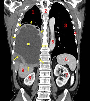
CT scan of a patient with mesothelioma,
coronal section (the section follows the plane the divides the body in a front and a back half). The mesothelioma is indicated by yellow arrows, the central
pleural effusion (fluid collection) is marked with a yellow star. Red numbers: (1) right lung, (2) spine, (3) left lung, (4) ribs, (5)
descending part of the
aorta, (6)
spleen, (7) left
kidney, (8) right kidney, (9)
liver.
Diagnosing mesothelioma is often difficult, because the symptoms are similar to those of a number of other conditions. Diagnosis begins with a review of the patient's medical history. A history of exposure to asbestos may increase clinical suspicion for mesothelioma. A physical examination is performed, followed by chest X-ray and often lung function tests. The X-ray may reveal pleural thickening commonly seen after asbestos exposure and increases suspicion of mesothelioma. A CT (or CAT) scan or an MRI is usually performed. If a large amount of fluid is present, abnormal cells may be detected by cytology if this fluid is aspirated with a syringe. For pleural fluid this is done by a pleural tap or chest drain, in ascites with an paracentesis or ascitic drain and in a pericardial effusion with pericardiocentesis. While absence of malignant cells on cytology does not completely exclude mesothelioma, it makes it much more unlikely, especially if an alternative diagnosis can be made (e.g. tuberculosis, heart failure).
If cytology is positive or a plaque is regarded as suspicious, a biopsy is needed to confirm a diagnosis of mesothelioma. A doctor removes a sample of tissue for examination under a microscope by a pathologist. A biopsy may be done in different ways, depending on where the abnormal area is located. If the cancer is in the chest, the doctor may perform a thoracoscopy. In this procedure, the doctor makes a small cut through the chest wall and puts a thin, lighted tube called a thoracoscope into the chest between two ribs. Thoracoscopy allows the doctor to look inside the chest and obtain tissue samples.
If the cancer is in the abdomen, the doctor may perform a laparoscopy. To obtain tissue for examination, the doctor makes a small opening in the abdomen and inserts a special instrument into the abdominal cavity. If these procedures do not yield enough tissue, more extensive diagnostic surgery may be necessary.
Risk factors
Working with asbestos is the major risk factor for mesothelioma. Mesothelioma is now known to occur in those who are genetically pre-disposed to it. A history of asbestos exposure exists in almost all cases. However, mesothelioma has been reported in some individuals without any known exposure to asbestos. In rare cases, mesothelioma has also been associated with irradiation, intrapleural thorium dioxide (Thorotrast), and inhalation of other fibrous silicates, such as erionite.
Asbestos is the name of a group of minerals that occur naturally as masses of strong, flexible fibers that can be separated into thin threads and woven. Asbestos has been widely used in many industrial products, including cement, brake linings, roof shingles, flooring products, textiles, and insulation. If tiny asbestos particles float in the air, especially during the manufacturing process, they may be inhaled or swallowed, and can cause serious health problems. In addition to mesothelioma, exposure to asbestos increases the risk of lung cancer, asbestosis (a noncancerous, chronic lung ailment), and other cancers, such as those of the larynx and kidney.
The combination of smoking and asbestos exposure significantly increases a person's risk of developing cancer of the airways (lung cancer, bronchial carcinoma). The Kent brand of cigarettes used asbestos in its filters for the first few years of production in the 1950s and some cases of mesothelioma have resulted. Smoking modern cigarettes does not appear to increase the risk of mesothelioma.
Some studies suggest that simian virus 40 (SV40) may act as a cofactor in the development of mesothelioma.
Treatment
Treatment of malignant mesothelioma using conventional therapies has not proved successful and patients have a median survival time of 6 - 12 months after presentation[citation needed]. The clinical behaviour of the malignancy is affected by several factors including the continuous mesothelial surface of the pleural cavity which favours local metastasis via exfoliated cells, invasion to underlying tissue and other organs within the pleural cavity, and the extremely long latency period between asbestos exposure and development of the disease.
Surgery
Surgery, either by itself or used in combination with pre- and post-operative adjuvant therapies, has proved disappointing. A pleurectomy/decortication is the most common surgery, in which the lining of the chest is removed. Less common is an extrapleural pneumonectomy (EPP), in which the lung, lining of the inside of the chest, the hemi-diaphragm and the pericardium are removed. It is not possible to remove the entire mesothelium without killing the patient.


